
The importance of functional validation in cryopreservation research
Standard viability assays are the equivalent of checking if a car has all four tires and then declaring it ready for a Formula 1 race. It might not have an engine, but at least the tires aren't flat.

In the world of cryopreservation research, we often fall in love with the “still life.” We produce stunning electron microscopy showing perfectly preserved organelles, or very high cell viability outcomes. We immediately go and publish that as “an improved cryopreservation protocol”. But in the quest for true biological stasis, there is a dangerous gap between structural preservation and biological utility.
To move the needle on organ banking, and eventual whole organism POC, we must prioritize functional validation: the proof that a biological system doesn’t just look “fine,” but can still perform its job. Cryopreservation has reached the point where structural validation is no longer an adequate metric of progress; functional performance must be the new gold standard.
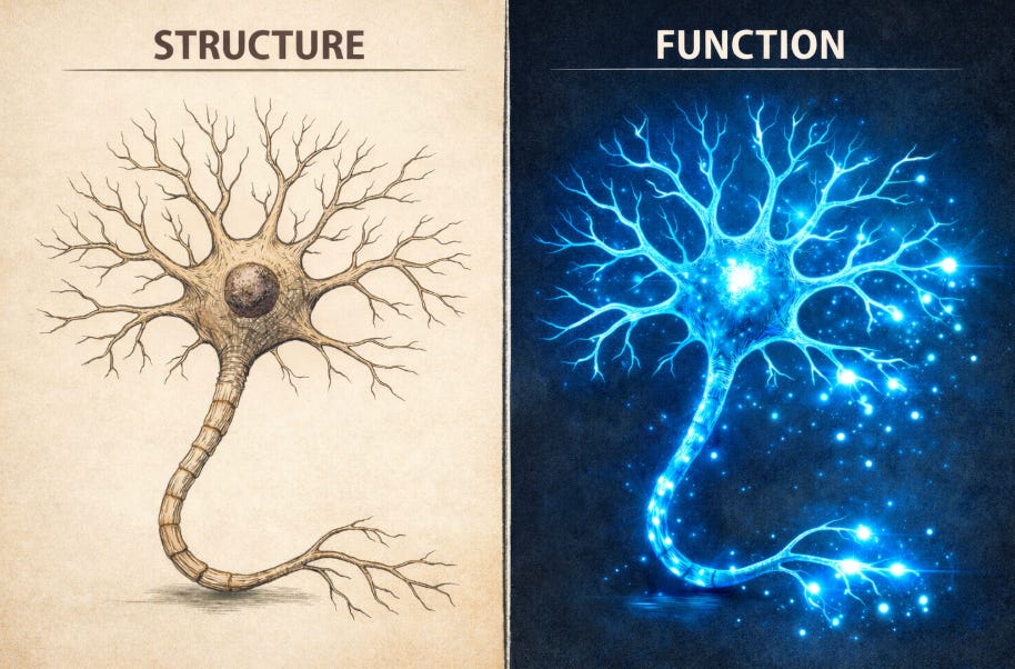
Structural vs. Functional
Structural validation tells us the “parts” are there. Functional validation tells us the “machine” still runs. In neurobiology, for instance, a vitrified and rewarmed slice of the hippocampus might show intact synaptic densities under a microscope. However, without electrophysiological testing such as Long-Term Potentiation (LTP) protocols, we cannot claim the circuit is “preserved.” If the neurons cannot fire or maintain a resting membrane potential, we haven’t preserved a brain; we’ve preserved a very complex sculpture.
To check the effectiveness of their protocols (That surprisingly often still operate by manipulating the ratios of the 4 horseman: DMSO, EG, PG and sucrose/trehalose), most groups default to immunofluorescence, cell counts, or Western blots because they are cheap, fast, and operationally simple. A full round of IF or a Western blot costs well under $50–$150 per sample in consumables, requires only a microscope or a gel rig that every lab already owns, and can be completed within a day. These assays scale easily and fit into the workflow of junior technicians.
Functional assays, in contrast, are not prohibitively expensive, but they do require different infrastructure, expertise, and time. Of course, they are tissue-specific, but some examples include:
Enzyme activity assays:
Many kits run $3–20 per reaction;
even kinetic assays on plate readers cost less than an antibody.Live-cell imaging of calcium flux, membrane potential, or synaptic activity:
Dyes are $5–10 per sample,
time on a decent confocal ~$20–80/hr internal recharge rate.
Out of assays that require a one time big investment but then serve for years:
Mitochondrial respiration (Seahorse XF):
Approx. $45–70 per well for sensor cartridges + plates;
initial instrument cost $60–130k, so I get that some labs might not have it
runtime ~2–3 hours.
Once the instrument exists, per-sample costs are comparable to a good antibody.Electrophysiology (patch-clamp, MEA):
Big up-front cost ($40–150k),
but per-sample consumables are low (~$5–15).
The true cost is expertise and labor, not materials.Flow cytometry (functional reporter dyes):
Per-sample consumables $2–6 per sample for live-cell functional dyes (calcium flux, ROS, mitochondrial potential, apoptosis), plus basic tubes and buffers.
The initial instrument cost is high ($80–350k, depending on configuration), so not every lab owns one directly, but almost all institutions have a shared core facility.
These were for cells. For organs it goes without saying that the ultimate success test is transplantation.
In other words, the barrier is not money, it’s culture and convenience (and the specialized trainee to run eg patch clamping, but that is case-specific)
For most experiments, running a functional readout costs:
2–3× more per sample than a basic Western blot,
but 10× more information about whether the cell actually works.
The difference between “cheap” and “functional” is typically a few dozen dollars per sample, not thousands (assuming the lab has access to equipment (or collaborators)). Cryopreservation research has historically chosen structural assays because they’re accessible and familiar, not because functional assays are financially out of reach.
Do you have any other '“cheap and easy but useful” functional assay examples?
Why Validation Matters On Many Levels
True functional validation must happen across every layer of biological organization:
Cellular Level: Cellular “viability” assays like Trypan Blue exclusion, Calcein-AM or DAPI staining set an exceptionally low bar. They report membrane integrity or general reductive activity, but say little about whether the cell can perform the function it is biologically designed for (and are so easy to misinterpret depending on what resolution you set, or which counting paramters you use). Across cell types, cryopreservation routinely preserves morphology and markers while degrading performance. Cardiomyocytes may remain viable yet lose contractility or exhibit abnormal excitation–contraction coupling. Immune cells frequently maintain surface markers and transcriptomes but show reduced proliferation, cytokine release, phagocytosis, migration, or cytotoxicity. Neural cells can look morphologically intact yet lose calcium synchrony, axonal transport efficiency, neurotransmission fidelity, or spontaneous electrophysiological activity.
The implication is straightforward: meaningful post-thaw assessment must evaluate contractility, metabolic flux, chemotaxis, electrophysiology, enzyme kinetics, or calcium signaling, not simply membrane integrity or marker expression.
Organ Level: At the organ scale, functional validation is non-negotiable because structure and physiology frequently diverge in meaningful ways after cryopreservation or traditional preservation methods. Simply observing intact vasculature or acceptable histology does not guarantee an organ will perform its biological functions once rewarmed or reperfused.
For instance, normothermic machine perfusion (NMP) has emerged clinically because static cold storage alone often fails to sustain metabolic and functional integrity in donor organs. NMP enables ongoing metabolism and direct functional evaluation including oxygen consumption, lactate clearance, bile production, and urine output during ex vivo perfusion, and these metrics correlate more strongly with graft viability than histology alone. This phenomenon has been repeatedly documented across organs such as liver and kidney grafts subjected to NMP prior to transplantation.
In kidney transplantation research, ex vivo perfusion parameters such as creatinine clearance, arterial flow patterns, and oxygen consumption during normothermic perfusion have been used to distinguish functional from non-functional grafts, even when biopsy histology shows comparable architecture. A detailed cryopreservation study of vitrified kidneys (Han et al., 2023 - if you are still not familiar with it, shame on you;)) further demonstrates this point empirically: histology and ultrastructure appeared near-normal, yet functional recovery measured by urine production, venous and urine electrolyte composition, and metabolic activity during NMP was essential to establish true viability. After transplantation into nephrectomized recipients, these functional measures normalized over time despite only focal histological injury, underlining the fact that morphology alone cannot predict systemic performance.
Similarly, in liver preservation, clinical and preclinical data show that dynamic perfusion metrics such as lactate clearance, bile output, and ATP content during NMP are far more predictive of transplantability than static histological assessments. Whereas static cold preservation may maintain gross structure, it often permits metabolic deterioration and ischemia–reperfusion injury that only becomes evident through dynamic function testing.
Even outside strict cryopreservation work, transplantation research emphasizes that functional testing on machine perfusion platforms, measuring vascular resistance, metabolic waste clearance, oxygen extraction, and organ-specific secretory function, outperforms histology and conventional viability staining in predicting outcomes. This reflects a broader consensus in the field that structure is a necessary but insufficient indicator of biological utility.
The Path Forward: Standardization
If we want cryopreservation to be a scalable pillar of regenerative medicine, we need a “Functional Registry.” We must move beyond “post-thaw recovery” percentages and toward standardized benchmarks:
If you insist on cell research, do at least a metabolic flux analysis: Metabolic flux analysis is the most universally informative functional metric for cryopreserved cells, because energy metabolism is the earliest and most sensitive point of failure. However, it should be treated as a baseline assay rather than the definitive functional test, since optimal readouts are cell-type–specific (e.g., electrophysiology for neurons, contractility for cardiomyocytes, cytotoxicity for immune cells).
Integrated “Omics”: A persistent limitation in cryopreservation research is the near-total absence of post-thaw multi-omics profiling. When a functional assay under-performs, each omics layer supplies a distinct forensic tool:
Transcriptomics immediately flags whether the problem is upstream at the gene-expression level: it reveals stress-induced transcriptional programs (e.g., massive upregulation of HSPA1A, ATF4, or CHOP), repression of lineage-specific or functional genes (e.g., downregulation of INS, PDX1, or MYH6), or aberrant activation of apoptosis/senescence modules. If the cells look alive but do not function, transcriptomics tells you the blueprint itself is corrupted.
Proteomics localizes damage one layer downstream: it quantifies actual protein abundance, post-translational modifications, misfolding (increased ubiquitinated or carbonylated proteins), chaperone overload (elevated HSP70/90), or proteolytic degradation (cleaved caspases, proteasome subunits). It can show that even though the mRNA for a critical enzyme or channel is present, the functional protein is missing, aggregated, or inactivated—explaining why the functional test collapsed.
Metabolomics (and lipidomics) exposes the final functional bottleneck: it detects ATP/ADP collapse, accumulation of reactive oxygen species markers (8-OHdG, 4-HNE), depleted glycolytic or TCA-cycle intermediates, membrane lipid peroxidation, or toxic cryoprotectant metabolites. These profiles reveal whether the cell has entered a metabolically crippled state even though transcription and translation initially occurred—directly accounting for loss of contractility, secretion, or motility in the functional assay.
Through integrating these readouts, researchers can now answer the precise question “why did the functional test fail?” with mechanistic insights: transcriptional repression, protein-level damage, or metabolic collapse. This granularity transforms cryopreservation optimization from trial-and-error into targeted engineering of the exact step that broke.
In Vivo Transplantation: The ultimate functional test: can a preserved tissue integrate and sustain life in a model organism? It can also apply to organoids (see my latest post for examples)
Why This Matters More Than Ever
Reproducibility and Scalability: Cryoexperiments are expensive and technically demanding. If protocols are validated only on viability, labs waste years chasing artifacts. Rigorous functional benchmarks let us compare protocols apples-to-apples and accelerate iteration.
Regulatory and Clinical Reality: Regulators (FDA, EMA) demand potency assays for advanced therapy medicinal products (ATMPs). Viability alone won’t cut it for organ banking or future cryonics-derived regenerative therapies.
AI Scaling: At the intersection of AI and cryopreservation, functional data is gold. Based on the results of multiple functional assays, AI can predict optimal vitrification cocktails, cooling and warming protocols, and foundational models simulate rewarming damage. But the training data must be functional metrics, not just survival. Garbage viability data in, garbage predictions out.